


Every year in March, the world observes World Kidney Day – an initiative dedicated to raising awareness about kidney health and the importance of early prevention.
In 2026 World Kidney Day will be marked on March 12 and serves as a reminder of how important it is to take care of these vital organs.
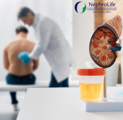
The kidneys play a key role in the human body – they filter waste products from the blood, regulate fluid and electrolyte balance, help control blood pressure, and participate in hormone production. Despite their essential functions, kidney diseases often develop without clear symptoms in their early stages, which makes prevention extremely important.
One of the most effective ways to prevent kidney disease is maintaining a healthy lifestyle. A balanced diet with less salt and fewer processed foods, adequate water intake, regular physical activity, and maintaining a healthy body weight are among the main factors supporting good kidney function. Controlling blood pressure and blood sugar levels is also crucial, as hypertension and diabetes are among the leading causes of chronic kidney disease.
Regular preventive check-ups can detect problems at an early stage.
The main blood tests that indicate kidney function include several laboratory markers through which doctors assess how well the kidneys filter the blood.
1. Creatinine (Serum Creatinine)
This is the most commonly used indicator for assessing kidney function.
Creatinine is a waste product of muscle metabolism that is normally excreted by the kidneys.
Elevated levels may indicate reduced kidney function.
It is also used to calculate another important indicator – eGFR.
2. eGFR (estimated Glomerular Filtration Rate)
This is the most accurate indicator of actual kidney function.
It is calculated based on:
-
creatinine
-
age
-
sex
-
sometimes body weight
It shows how many milliliters of blood the kidneys filter per minute. A decreased value may be a sign of chronic kidney disease.
3. Urea (Blood Urea Nitrogen – BUN)
Urea is a waste product formed during the breakdown of proteins.
When the kidneys are not functioning properly, the level of urea in the blood increases.
This indicator is usually evaluated together with creatinine.
4. Uric Acid
The kidneys play a role in eliminating it from the body.
Elevated levels may be associated with:
-
kidney problems
-
gout
-
metabolic disorders.
5. Electrolytes (sodium, potassium, chlorides)
The kidneys regulate electrolyte balance.
When kidney function is impaired, the following may occur:
-
increased potassium
-
changes in sodium and chloride levels.
Additional test
In addition to blood tests, a urine test (albumin/protein in urine) is almost always performed, which is very important for the early detection of kidney damage.
World Kidney Day is an opportunity to remind ourselves that kidney health largely depends on our daily choices. Increasing public awareness, early diagnosis, and active prevention can significantly reduce the risk of kidney diseases and improve the quality of life for millions of people.